|
Overview
 Fairly common in infants and small children, but also prevalent in adults, flat feet becomes a problem if pain or discomfort is present in the foot or even around the knee and lower leg area. Pain around the knee and lower leg areas can arise because flat feet can alter proper foot and leg alignment, which will put unusual strain on the knee. Adults can develop the condition as the result of injury or even from normal wear and tear due to the natural aging process. Causes Flat feet can be caused by injury, aging, and weight gain. They can cause pain in the feet and may lead to pain in other parts of the body such as the ankles, knees, or hips. For this reason, it behooves us to treat fallen arches. The question becomes how to do so. Symptoms Flat feet may not cause any symptoms at all. Rigid flat feet may cause pain, calluses, blisters, or skin redness on the inner side of the foot. A stiff foot, weakness or numbness of the foot, Rapid wearing out of shoes-worn shoes lean in toward each other. Difficulty or pain with activities like running-in the foot, knee or hip. Diagnosis Your doctor will ask about your symptoms and medical history. A physical and foot exam will be done. Flat feet can be diagnosed by appearance. To determine if the foot is rigid, you may be asked to do some simple tasks. pes cavus Non Surgical Treatment Some patients with flat feet may automatically align their limbs in such a way that unpleasant symptoms never develop. In such cases treatment is not usually required. Pain in the foot that is caused by flat feet may be alleviated if the patient wears supportive well-fitted shoes. Some patients say that symptoms improve with extra-wide fitting shoes. Fitted insoles or orthotics (custom-designed arch supports) may relieve pressure from the arch and reduce pain if the patient's feet roll or over-pronate. The benefits of an orthotic only exist while it is being worn. Patients with tendonitis of the posterior tibial tendon may benefit if a wedge is inserted along the inside edge of the orthotic - this takes some of the load off the tendon tissue. Wearing an ankle brace may help patients with posterior tibial tendinitis, until the inflammation comes down. Rest, doctors may advise some patients to rest and avoid activities which may make the foot (feet) feel worse, until the foot (feet) feels better. A combination of an insole and some kind of painkiller may help patients with a ruptured tendon, as well as those with arthritis. Patients with a ruptured tendon or arthritis who find insoles with painkillers ineffective may require surgical intervention. Patients, usually children, whose bones did not or are not developing properly, resulting in flat feet from birth, may require surgical intervention to separate fused bones (rare). Bodyweight management, if the patient is obese the doctor may advise him/her to lose weight. A significant number of obese patients with flat feet who successfully lose weight experience considerable improvement of symptoms. Surgical Treatment 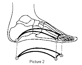 Surgery for flat feet is separated into three kinds: soft tissue procedures, bone cuts, and bone fusions. Depending on the severity of the flat foot, a person?s age, and whether or not the foot is stiff determines just how the foot can be fixed. In most cases a combination of procedures are performed. With flexible flat feet, surgery is geared at maintaining the motion of the foot and recreating the arch. Commonly this may involve tendon repairs along the inside of the foot to reinforce the main tendon that lifts the arch. When the bone collapse is significant, bone procedures are included to physically rebuild the arch, and realign the heel. The presence of bunions with flat feet is often contributing to the collapse and in most situations requires correction. With rigid flat feet, surgery is focused on restoring the shape of the foot through procedures that eliminate motion. In this case, motion does not exist pre-operatively, so realigning the foot is of utmost importance. The exception, are rigid flat feet due to tarsal coalition (fused segment of bone) in the back of the foot where freeing the blockage can restore function. Prevention Strap the arches into the anatomically correct positions with athletic tape and leave them like this for some time. If the fallen arches are an issue with the muscular structure, this may give the muscles an opportunity to strengthen. This is definitely not a fallen arches cure all the time but it can help prevent it more times than not. Ask a doctor or physical therapists to show you how to do this taping. Find shoes that fit. This may require that you get your foot measured and molded to ensure that the shoe will fit. Shoes that are too big, too tight or too short, may not directly cause the fallen arches, but they can assist with the damage to the area. These shoes should have thick cushioning inside and have plenty of room for your toes. Walk without shoes as much as possible. Shoes directly assist with weakening and distorting the arches of the feet so going without shoes can actually help strengthen your arches and prevent fallen arches. Walking on hard and bumpy surfaces barefooted makes the muscles in your feet strengthen in order to prevent injury. It is a coping mechanism by your body. Insert heel cups or insoles into the shoes that you wear the most. Many people wear uncomfortable shoes to work and these are the same shoes that cause their arches the most problems. Inserting the heel cups and insoles into these shoes can prevent fallen arches from occurring. Many people place these inserts into all their shoes to ensure support. Ask a medical professional, either your doctor or a physical therapist, about daily foot exercises that may keep the arches stronger than normal. Many times, you can find exercises and stretches on the Internet on various websites. Curling your toes tightly and rotating your feet will help strengthen your longitudinal arches. Relax your feet and shake them for a minute or so before you do any arch exercises. This will loosen the muscles in your feet that stay tight due to normal daily activities. Wear rigid soled sandals whenever possible to provide a strong support for your arches. Wooden soled sandals are the best ones if available. Walk or jog on concrete as much as you can. This will create a sturdy support for your arches. Running or walking in sandy areas or even on a treadmill, does not give rigid support. Instead, these surfaces absorb the step, offering no support for arches. After Care Patients may go home the day of surgery or they may require an overnight hospital stay. The leg will be placed in a splint or cast and should be kept elevated for the first two weeks. At that point, sutures are removed. A new cast or a removable boot is then placed. It is important that patients do not put any weight on the corrected foot for six to eight weeks following the operation. Patients may begin bearing weight at eight weeks and usually progress to full weightbearing by 10 to 12 weeks. For some patients, weightbearing requires additional time. After 12 weeks, patients commonly can transition to wearing a shoe. Inserts and ankle braces are often used. Physical therapy may be recommended. There are complications that relate to surgery in general. These include the risks associated with anesthesia, infection, damage to nerves and blood vessels, and bleeding or blood clots. Complications following flatfoot surgery may include wound breakdown or nonunion (incomplete healing of the bones). These complications often can be prevented with proper wound care and rehabilitation. Occasionally, patients may notice some discomfort due to prominent hardware. Removal of hardware can be done at a later time if this is an issue. The overall complication rates for flatfoot surgery are low.
0 Comments
Overview
 Heel pain is a common foot condition. It's usually felt as an intense pain when using the affected heel. Heel pain usually builds up gradually and gets worse over time. The pain is often severe and occurs when you place weight on the heel. In most cases, only one heel is affected, although estimates suggest that around a third of people have pain in both heels. The pain is usually worse first thing in the morning, or when you first take a step after a period of inactivity. Walking usually improves the pain, but it often gets worse again after walking or standing for a long time. Some people may limp or develop an abnormal walking style as they try to avoid placing weight on the affected heel. Causes Heel pain is not usually caused by a single injury, such as a twist or fall, but rather the result of repetitive stress and pounding of the heel. The most common causes of heel pain are Plantar fasciitis (plantar fasciosis) - inflammation of the plantar fascia. The plantar fascia is a strong bowstring-like ligament that runs from the calcaneum (heel bone) to the tip of the foot. When the plantar fasciitis is stretched too far its soft tissue fibers become inflamed, usually where it attaches to the heel bone. Sometimes the problem may occur in the middle of the foot. The patient experiences pain under the foot, especially after long periods of rest. Some patients have calf-muscle cramps if the Achilles tendon tightens too. Heel bursitis, inflammation of the back of the heel, the bursa (a fibrous sac full of fluid). Can be caused by landing awkwardly or hard on the heels. Can also be caused by pressure from footwear. Pain is typically felt either deep inside the heel or at the back of the heel. Sometimes the Achilles tendon may swell. As the day progresses the pain usually gets worse. Heel bumps (pump bumps) common in teenagers. The heel bone is not yet fully mature and rubs excessively, resulting in the formation of too much bone. Often caused by having a flat foot. Among females can be caused by starting to wear high heels before the bone is fully mature Tarsal tunnel syndrome, a large nerve in the back of the foot becomes pinched, or entrapped (compressed). This is a type of compression neuropathy that can occur either in the ankle or foot. Chronic inflammation of the heel pad, caused either by the heel pad becoming too thin, or heavy footsteps. Stress fracture, this is a fracture caused by repetitive stress, commonly caused by strenuous exercise, sports or heavy manual work. Runners are particularly prone to stress fracture in the metatarsal bones of the foot. Can also be caused by osteoporosis. Severs disease (calcaneal apophysitis) the most common cause of heel pain in child/teenage athletes, caused by overuse and repetitive microtrauma of the growth plates of the calcaneus (heel bone). Children aged from 7-15 are most commonly affected. Achilles tendonosis (degenerative tendinopathy) also referred to as tendonitis, tendinosis and tendinopathy. A chronic (long-term) condition associated with the progressive degeneration of the Achilles tendon. Sometimes the Achilles tendon does not function properly because of multiple, minor microscopic tears of the tendon, which cannot heal and repair itself correctly, the Achilles tendon receives more tension than it can cope with and microscopic tears develop. Eventually, the tendon thickens, weakens and becomes painful. Symptoms Sever?s Disease. This is a condition that occurs in 10 - 15 year old children, predominantly boys and is associated with running and repetitive jumping. It is also associated with flimsy footwear that kids may wear. It occurs when the Achilles tendon continually pulls on the apophysis of the calcaneum and does not allow for it to fuse with the body of the calcaneum. Calcaneal enthesopathy. This occurs when there is repetitive trauma at the attachment of the Achilles tendon, resulting in a spur from the calcaneum up into the Achilles tendon. It is usually visualized on x-ray and may be tender if there is an associated bursitis or tendonitis. "Pump Bump". Also known as Haglund?s Deformity, this is a bony enlargement that exists on the back of the heel - usually related to a congenital abnormality or with chronic bursitis, causing a thickening. There may have already been trauma or pressure from footwear. Treatment is usually protection of the bump and correct footwear. Associated with a symmetrical swelling at the base of the Achilles tendon. It is usually related to repetitive trauma or inappropriate footwear. It is often red and hot in the early stages. Treatment is usually to correct the footwear, provide padding and treat the local symptoms e.g. ice, rest, physiotherapy and cortisone injection. Fat Pad Syndrome. Direct contact with the base of the heel may result in trauma to the fat pad. Related to obesity, training on hard surfaces, uneven grounds, poor shoes especially overlarge shoes which can cause shearing forces on the heel. These conditions are renowned for taking a long time to recover - usually many months. Diagnosis In most cases, your GP or a podiatrist (a specialist in foot problems and foot care) should be able to diagnose the cause of your heel pain by asking about your symptoms and medical history, examining your heel and foot. Non Surgical Treatment The proper treatment for your heel pain depends entirely on the specific cause(s) of your symptoms. Therefore, it is critical to understand the cause(s) of your symptoms before beginning any treatment program and if you are unsure, then seeking medical advice is essential to develop the proper treatment program for your condition. Some common treatments are listed and can be performed at home. Keep in mind that not all of these treatments are appropriate for every condition, but they usually a good place to start. Rest, reducing activities for a few days can help to reduce the most severe pain. Ice, applying ice to the heel for 10 minutes several times a day will help to reduce inflammation. Stretching exercises, to lengthen the muscles in the back of the leg, including the hamstrings, will help to ease pain, reduce focal pressures to your feet and assist in recovery. For plantar fasciitis, this may be the best treatment of all. Avoid going barefoot, when without shoes excessive stress and strain is placed on the plantar fascia. Proper shoe gear, supportive shoes that fit and are not too worn along with good arch support help to reduce the stress and strain on the plantar fascia over time. Medications, non-steroidal anti-inflammatory medication, such as Motrin (ibuprofen), may help to reduce inflammation. If the pain persists or worsens after a couple of days, an appointment may be necessary where Dr. Talarico may add one or more of these additional modalities to your treatment program. Orthotic b, whether pre-fabricated or custom orthotic is used, these devices can help reduce the underlying structural abnormalities of the foot which have lead to the development of plantar fasciitis. These are often used to limit the recurrence of plantar fasciitis pain. Strapping, a special taping technique to help reduce the strain on the fascia. Injection therapy, in some instances injections are used to reduce the inflammation and reduce pain. Night Splint, this allows you to maintain an extended stretch on the plantar fascia while sleeping. Over time, this has shown to reduce the morning pain which some people experience. Removable Walking Cast, in some case of severe heel pain this may be used to keep your foot immobile for a few weeks allowing it to rest and heal. Physical Therapy may be recommended to aid in pain relief. At The Foot & Ankle Center, PC, Dr Talarico will often utilize two additional in-office modalities, EPAT and MLS Laser Therapy, which are very effective in treating most inflammatory conditions of the foot and ankle, including plantar fasciitis. Surgical Treatment Extracorporeal shockwave therapy (EST) is a fairly new type of non-invasive treatment. Non-invasive means it does not involve making cuts into your body. EST involves using a device to deliver high-energy soundwaves into your heel. The soundwaves can sometimes cause pain, so a local anaesthetic may be used to numb your heel. It is claimed that EST works in two ways. It is thought to have a "numbing" effect on the nerves that transmit pain signals to your brain, help stimulate and speed up the healing process. However, these claims have not yet been definitively proven. The National Institute for Health and Care Excellence (NICE) has issued guidance about the use of EST for treating plantar fasciitis. NICE states there are no concerns over the safety of EST, but there are uncertainties about how effective the procedure is for treating heel pain. Some studies have reported that EST is more effective than surgery and other non-surgical treatments, while other studies found the procedure to be no better than a placebo (sham treatment). no foot pain Prevention  Make sure you wear appropriate supportive shoes. Don't over-train in sports. Make sure you warm up, cool down and undertake an exercise regime that helps maintain flexibility. Manage your weight, obesity is a factor in causing plantar fasciitis. Avoid walking and running on hard surfaces if you are prone to pain. You should follow the recognized management protocol "RICED" rest, ice, compression, elevation and diagnosis. Rest, keep off the injured ankle as much as possible. Ice, applied for 20 minutes at a time every hour as long as swelling persists. Compression, support the ankle and foot with a firmly (not tightly) wrapped elastic bandage. Elevation, keep foot above heart level to minimize bruising and swelling. Diagnosis. Consult a medical professional (such as a Podiatrist or doctor) especially if you are worried about the injury, or if the pain or swelling gets worse. If the pain or swelling has not gone down significantly within 48 hours, also seek treatment. An accurate diagnosis is essential for proper rehabilitation of moderate to severe injuries. |
AuthorWrite something about yourself. No need to be fancy, just an overview. Archives
July 2017
Categories |
 RSS Feed
